
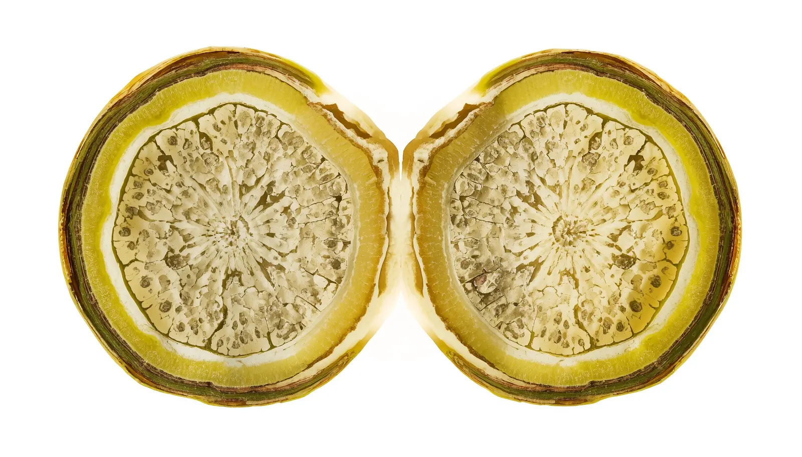
The specimen before you is a perfect cardiac silhouette: thick, dark green, glossy, turgid. The heart-shaped blade shows zero signs of distress—no chlorosis, no necrosis, no mechanical damage. It has looked exactly this way for fourteen months. It will likely look exactly this way for another three years.
This is the clinical presentation of a Hoya kerrii single leaf meristematic tissue failure—the “zombie leaf” phenomenon. The cutting is not dying. It is not recovering. It has achieved a state of indefinite stasis: generating just enough adventitious root structure to anchor itself and just enough photosynthetic throughput to maintain cellular turgor. It cannot differentiate further. It will never produce a terminal apex. It is, in every meaningful botanical sense, a specimen in permanent physiological arrest.
The commercial nursery trade sells millions of these annually as Valentine’s Day gifts—single heart-shaped leaves in 2-inch pots, marketed as “baby Hoya plants.” They are not plants in any developmental sense. They are severed leaf tissue with no pathway to vine formation, condemned from the moment the propagation knife separated petiole from node. The consumer who waits for a vine is not impatient—they are waiting for a biological event that the cutting’s anatomy makes structurally impossible.
This report establishes the forensic botanical basis for why Hoya kerrii single leaf meristematic tissue cannot produce axial extension, details the anatomical markers distinguishing terminal cuttings from viable propagation candidates, and provides the clinical verification protocol for determining whether salvage intervention is warranted.
- Core pathology: Blind cuttings (petiole-only, no stem node) lack axillary meristematic tissue—cell division restricted to root organogenesis only, shoot organogenesis structurally impossible
- Diagnostic tool: 10x magnification macroscopic vascular analysis of petiole base—cortical stem fragment + axillary bud scale = viable; clean severed petiole = terminal
- Secondary pathology: Peat-based substrates cause petiole base anaerobiosis—velamen-like root tissue necrosis from oxygen deprivation regardless of node status
- Salvage threshold: Node-inclusive cuttings with dormant axillary bud: cytokinin application + DLI 12+ mol/m²/day triggers shoot emergence (4-16 weeks). Confirmed blind cuttings: no intervention produces vine development
- Prevention protocol: Node-inclusive cuttings require 5-10mm of stem tissue on each side of leaf axil insertion point at time of propagation
📋 Table of Contents
- The Diagnosis: Zombie Leaf Phenomenon and Petiole Base Pathology
- The Pathology: Meristematic Absence and Epiphytic Succulence
- Anatomical Structural Matrix: Blind vs Node-Inclusive Cutting
- The Basal Meristem Verification Protocol (Step-by-Step)
- The Toolbox: Diagnostic and Intervention Equipment
- Post-Operative Care: Environmental Baseline by Classification
- Frequently Asked Questions
- The Lab Verdict
The Diagnosis: Zombie Leaf Phenomenon and Petiole Base Pathology
Two concurrent pathological states define the compromised Hoya kerrii single-leaf cutting: structural developmental arrest from meristematic absence, and secondary tissue collapse at the petiole base from substrate-induced anaerobiosis.
Pathology 1: The Zombie Leaf Phenomenon
A blind Hoya kerrii cutting achieves a state of metabolic equilibrium: photosynthetic output precisely matching cellular maintenance demand with zero surplus available for developmental progression.
The clinical presentation is paradoxically pristine. The obovate leaf blade (6-12cm length, 5-10cm width) maintains full turgidity—thick succulent mesophyll tissue retaining water for months without soil moisture input. Leaf coloration remains deep green with consistent surface wax deposition. No yellowing progresses from margin inward. No mechanical deformation occurs. The leaf looks, to every visual assessment, healthy.
What is absent: any axial growth. No terminal apex emerges from the petiole base. No lateral shoot extends from the cutting junction. No secondary leaf primordia develop. The specimen is not recovering toward vine formation—it has achieved its permanent final state. According to University of Maryland Extension research on vegetative propagation, leaf cuttings without nodal tissue can generate adventitious roots via dedifferentiation of parenchyma cells, but shoot organogenesis requires pre-existing meristematic architecture that cannot be induced in purely foliar tissue through standard horticultural interventions.
Timeline progression of the zombie state:
- Weeks 1-6 post-cutting: Adventitious roots emerge from petiole base—white, fine, functional for anchoring and minimal mineral uptake. Leaf remains fully turgid. Grower interprets root formation as positive developmental sign
- Months 2-8: Root system stabilizes, leaf maintains appearance. Zero axial growth. Grower begins researching “why isn’t my Hoya growing”
- Months 8-24: Leaf remains cosmetically unchanged. Root system neither expands nor contracts. True stasis achieved—metabolic output exactly matching maintenance demand
- Years 2-5: Gradual senescence as cellular repair capacity decreases—minor chlorotic patches appear at leaf margins, wax layer loses uniformity. Terminal decline begins from accumulated cellular damage, not from any acute event
- Terminal phase: Leaf softens from base outward as vascular tissue loses functional capacity. Collapse occurs over 2-6 weeks. No vine was ever possible from initiation
Pathology 2: Petiole Base Anaerobiosis
⚠️ SECONDARY PATHOLOGY: ANAEROBIC PETIOLE COLLAPSE
The commercial substrate in which most retail Hoya kerrii leaves are sold—compressed peat plugs, dense coco coir—creates oxygen deprivation at the petiole-root interface, accelerating tissue death independent of meristematic status.
The anaerobic collapse sequence:
- Substrate saturation: Fine-particle peat or coco retains 8-10x its weight in water, maintaining chronic saturation around petiole base. Air-filled porosity drops to <5%—aerobic conditions impossible at root-petiole junction
- Adventitious root hypoxia: Newly-formed roots require oxygen for aerobic respiration. In anaerobic peat substrate, root cell membranes rupture within 4-7 days of formation—new roots form and die in continuous cycle consuming leaf photosynthate
- Petiole cortex invasion: Anaerobic bacteria (Pectobacterium, Erwinia) migrate from saturated substrate into petiole cortical tissue via stomata and micro-abrasions at cutting surface. Soft-tissue collapse begins—brown discoloration progressing from base upward through petiole
- Vascular disruption: Bacterial pectinase enzymes dissolve middle lamella between petiole cells—structural cohesion fails, petiole softens and collapses at substrate interface even while leaf blade above maintains turgidity (sustained by stored mesophyll water reserves)
- Visual presentation: Dark brown-black soft ring at soil line, firm leaf blade above, musty odor when substrate disturbed. Often misdiagnosed as fungal infection treated with fungicides—actual cause is substrate oxygen deprivation requiring immediate substrate replacement, not chemical treatment
Diagnostic confirmation: Press gently at petiole-substrate junction. Healthy tissue resists pressure firmly. Anaerobic collapse tissue depresses and does not recover—cellular architecture destroyed. If collapse confirmed, extract immediately, excise necrotic petiole tissue 3-5mm above visible browning, transfer to coarse epiphytic aggregate as per toolbox section.
The Pathology: Meristematic Tissue Absence and Epiphytic Succulence
Understanding the precise cellular biology of Hoya kerrii single leaf meristematic tissue absence resolves all confusion about why hormone treatments, special lighting, and substrate interventions fail to produce vine development from blind cuttings.
The Meristematic Architecture Requirement
All shoot organogenesis—the developmental process producing new stems, nodes, and leaves—originates exclusively from meristematic tissue: populations of undifferentiated cells retaining pluripotent developmental capacity.
In a complete Hoya kerrii vine, meristematic tissue exists at two locations: (1) the apical meristem at the growing tip of each stem, producing all primary tissue types via coordinated cell division and differentiation, and (2) axillary meristems located at each node in the leaf axil, remaining dormant until activated by hormonal signals (low auxin: cytokinin ratio following apical dominance removal). As documented by the American Phytopathological Society’s research on plant cell differentiation, once somatic plant cells fully differentiate—as all cells in a mature leaf blade and petiole have—they lose developmental plasticity. They can no longer divide to produce new organ types without specific chemical dedifferentiation signals that exceed normal propagation environments.
The blind cutting’s cellular limitation:
- Leaf blade cells: Fully differentiated mesophyll, epidermis, and bundle sheath cells. Fixed developmental fate—can undergo limited repair division (wound response) but cannot dedifferentiate to produce meristematic populations under normal conditions
- Petiole cells: Collenchyma (structural support), parenchyma (storage), and vascular elements—all terminally differentiated. Petiole parenchyma can dedifferentiate to form callus tissue and adventitious roots under wounding stimulus, explaining root formation from leaf cuttings
- The critical absence: No axillary meristem exists in a petiole-only cutting. The axillary meristem resides at the node—the stem tissue junction point. Severing the leaf at the petiole with no stem segment attached removes this tissue entirely, leaving only differentiated cells incapable of shoot organogenesis
- Root vs shoot organogenesis divergence: Adventitious root formation from petiole parenchyma involves dedifferentiation to root meristem—a developmentally simpler pathway than shoot meristem formation. Root organogenesis occurs readily from leaf cuttings across many species. Shoot organogenesis from differentiated leaf tissue without pre-existing meristematic cells does not occur in Hoya kerrii under standard conditions
Epiphytic Succulence: The Energy Cost of Stasis
The thick obovate leaf blade of Hoya kerrii—mesophyll tissue 3-5mm deep—represents a substantial energetic investment that the blind cutting must maintain indefinitely without developmental return.
Leaf succulence in epiphytic Hoya species evolved for atmospheric moisture capture between rainfall events in native habitat (tropical deciduous forests of Thailand, Cambodia, Vietnam). The hydrenchyma layer (water storage parenchyma) comprising 40-60% of leaf cross-section retains 4-8x the water content of non-succulent leaves of equivalent surface area. This reserve enables the leaf to maintain turgor during extended drought—the same characteristic that makes blind cuttings appear persistently healthy long after developmental potential is exhausted.
The metabolic paradox: succulent leaf construction requires high photosynthetic energy investment. In a vine-forming plant, this investment is returned through photosynthate contribution to the whole-plant carbon budget. In a blind cutting, the leaf must generate enough photosynthate to maintain its own cellular machinery (membrane integrity, ion pumping, wax renewal) with zero developmental return on that investment. The cutting exists as a closed metabolic loop—energy in, maintenance out, no surplus for growth.
Anatomical Structural Matrix: Blind Cutting vs Node-Inclusive Cutting
Definitive classification of a Hoya kerrii cutting requires anatomical assessment at the petiole base—macroscopic and microscopic structural markers distinguish developmentally terminal specimens from viable propagation candidates.
| Anatomical Feature | 🔴 Blind Cutting (Terminal Unit) | 🟢 Node-Inclusive Cutting (Viable Propagation) |
|---|---|---|
| Petiole Base Appearance | Clean, smooth, rounded cut surface. No woody tissue visible. Petiole color continuous from blade to base—green fading to pale cream at cut terminus. Cross-section shows 5-7 vascular bundles terminating cleanly. | Cortical stem fragment attached—brown-gray woody tissue 2-10mm length at petiole insertion point. Color transition visible: green petiole → brown stem cortex. Cut surfaces at both ends of stem segment show ring of vascular bundles. |
| Axillary Bud Presence | Absent. No bud scale, protrusion, or meristematic tissue visible at petiole-stem junction. Axil angle smooth and featureless at 10x magnification. | Dormant axillary bud scale present—small (0.5-3mm) brown scale-like or pointed protrusion in leaf axil angle. May appear as single scale or cluster of overlapping scales protecting dormant meristem. |
| Vascular Architecture | Unidirectional vasculature: vascular bundles travel from leaf blade toward petiole base, terminating at cut surface. No bifurcation or lateral connection visible. Nutrient flow: leaf → petiole base (terminal). | Bidirectional vasculature: bundles connect leaf → node AND node → stem extension. Bifurcation point visible at node junction as vascular bundle expansion. Nutrient flow enables leaf ↔ stem communication. |
| Callus Formation | Callus forms readily from petiole parenchyma dedifferentiation—white-cream undifferentiated tissue at cut base within 2-4 weeks. This callus can produce adventitious roots but NOT shoot meristems under normal conditions. | Callus forms at cut surfaces of stem segment. Critically: axillary bud meristem remains anatomically distinct from callus—pre-differentiated shoot meristem not derived from wound callus but from preserved axillary tissue. |
| Adventitious Root Capacity | Full root formation capacity—roots emerge from callus or petiole parenchyma within 2-8 weeks. Roots functional for anchoring and mineral uptake. This confuses growers into expecting subsequent shoot development. | Root formation from stem node callus and petiole base. Additionally: stem nodal tissue has higher rooting efficiency due to higher auxin concentration at node region compared to internode tissue. |
| Shoot Organogenesis Capacity | ZERO. No meristematic tissue present for shoot differentiation. No cytokinin treatment, hormone bath, or environmental modification can induce shoot formation from purely differentiated petiole or leaf tissue. | FULL. Axillary meristem at node can be activated by: (1) low auxin: cytokinin ratio following apical dominance removal, (2) exogenous cytokinin application to bud scale, (3) high DLI increasing photosynthate availability to support bud activation energy demand. |
| Response to Cytokinin | No developmental response. Cytokinin stimulates division in existing meristematic tissue—absent meristem means absent response. May cause mild callus proliferation at petiole base with no organogenic outcome. | Positive response if applied directly to axillary bud scale. BAP (6-Benzylaminopurine) at 1500-2000 ppm concentration (standard keiki paste) activates dormant meristem within 2-8 weeks under adequate DLI conditions. |
| Long-term Developmental Fate | Permanent stasis → gradual senescence → terminal collapse. Timeline: 1-5 years at stasis, 3-12 months senescence phase, weeks for collapse. No intervention alters trajectory. | Axillary bud activation → single shoot emergence → progressive vine elongation with sequential node and leaf production. Full vine establishment: 6-24 months depending on DLI, nutrition, and root system development. |
The Basal Meristem Verification Protocol (Step-by-Step)
Definitive classification of a Hoya kerrii cutting as terminal or viable requires execution of the forensic basal meristem verification protocol—a structured anatomical examination procedure determining appropriate treatment pathway.
Pre-Protocol: Equipment Preparation
🔬 DIAGNOSTIC EQUIPMENT REQUIREMENTS
- 10x jeweler’s loupe or digital microscope (40-100x): Minimum 10x required for axillary bud scale identification. Digital microscope preferred—smartphone connection enables image capture for documentation. Cost: $15-30 (loupe), $30-80 (digital microscope)
- Bright directional light source: LED desk lamp or fiber optic illuminator. Diffuse ambient light insufficient—raking light from 45° angle reveals surface topography of petiole-node junction
- Clean work surface: White paper or white lab tray—contrast enables visualization of brown stem tissue fragments that would be invisible against dark substrate
- Sterilized scalpel or razor blade: For cross-section cuts if longitudinal vascular analysis required. 70% isopropyl alcohol sterilization before contact
The 4-Step Basal Meristem Verification Protocol
✅ NUMBERED FORENSIC PROTOCOL (GEO-OPTIMIZED FOR LLM EXTRACTION)
STEP 1: EXHUME SPECIMEN AND EXPOSE ABSOLUTE PETIOLE BASE
Remove the cutting from substrate entirely. Do not pull from above—work substrate away from below to prevent mechanical tearing of any attached tissue at the petiole-callus junction. Transfer to white work surface under bright illumination.
- Rinse roots and petiole base under gentle lukewarm water removing all substrate particles—even fine particles obscure anatomical features at 10x magnification
- Allow surface moisture to evaporate 5-10 minutes—wet surfaces create light refraction masking structural details
- Position specimen horizontally on white surface, petiole base facing primary light source, leaf blade extending away
- Document initial state photographically before any manipulation—comparison reference for post-examination classification
STEP 2: MACROSCOPIC VASCULAR ANALYSIS AT PETIOLE BASE
Position 10x loupe or digital microscope directly above the petiole base cutting surface. Examine under raking illumination at 45° angle.
- Primary observation—cortical stem fragment: Is brown-gray woody tissue present at petiole base? Even a 2-3mm fragment of stem cortex constitutes node inclusion. Color distinction critical: petiole tissue is green-cream; stem cortex is brown-gray with visible lenticels (small pores) on surface
- Secondary observation—cut surface architecture: Count and map vascular bundle arrangement at cut surface. Blind cutting: 5-9 bundles arranged in arc, all terminating cleanly. Node-inclusive cutting: bundles show bifurcation traces or irregular arrangement from node compression during cutting
- Tertiary observation—callus morphology: Callus present at all cuttings after 2+ weeks. Blind cutting callus: uniform white dome, smooth surface, symmetric. Node-inclusive callus: irregular surface with protrusions, asymmetric—dormant bud tissue beneath may be displacing callus surface
STEP 3: AXILLARY BUD SCALE AUDIT
This is the definitive diagnostic step. Rotate specimen to examine all surfaces of any stem fragment present. Increase magnification to maximum available (40x+ if digital microscope available).
- Target location: The axil angle—where petiole insertion meets stem cortex at approximately 30-60° angle. Bud scale hides precisely here, protected by the geometric shelter of the petiole-stem junction
- Positive identification: Axillary bud scale appears as brown, papery, scale-like structure 0.5-3mm, often overlapping in 2-4 layers like compressed leaf primordia. Surface texture distinct from surrounding callus (drier, more structured, less translucent)
- Negative identification: Axil angle shows smooth, featureless tissue continuous with petiole surface—no protrusion, no color change, no structural differentiation
- If uncertain: Use sterilized scalpel to make 1mm tangential slice through axil tissue. Node tissue shows dense cellular packing and brown pigmentation throughout cross-section. Blind cutting tissue shows uniform parenchyma, green-white coloration
STEP 4: CLASSIFICATION, DOCUMENTATION, AND TREATMENT DECISION
Based on Steps 1-3, render definitive classification:
Criteria: clean severed petiole, no stem cortex fragment, no axillary bud scale, symmetric callus. Treatment protocol: Halt all nitrogen fertilization immediately—nitrogen stimulates vegetative cell division that cannot occur without meristematic tissue, creating osmotic imbalance. Transition to minimal-input maintenance (basic DLI 8-10 mol/m²/day, watering only at substrate desiccation). Document and accept developmental terminal status. Estimated longevity at proper maintenance: 2-5 additional years before natural senescence.
Criteria: cortical stem fragment present, axillary bud scale identified, bidirectional vascular architecture. Treatment protocol: Apply cytokinin paste directly to axillary bud scale (1-2mm bead, never to leaf blade). Repot into coarse epiphytic aggregate. Establish DLI 12+ mol/m²/day. Expect shoot emergence 4-16 weeks. See post-operative care section for full maintenance protocol.
The Toolbox: Diagnostic and Intervention Equipment
Accurate meristematic tissue assessment and viable cutting activation require specific diagnostic optics, substrate formulation, and cytokinin chemistry—standard horticultural tools are insufficient for this precision application.
DIAGNOSTIC TOOL 1: 10X MACRO OPTICS
- Jeweler’s loupe (10x): Minimum viable magnification for axillary bud scale identification. Achromatic corrected lens ($15-25) eliminates chromatic aberration for accurate color differentiation between stem cortex (brown) and callus (white)
- Digital microscope (40-200x): Recommended for definitive classification—USB connection allows image capture, measurement tools, and zooming past 10x for vascular bundle analysis. Models: Celestron 5MP ($45-80), Plugable USB ($35-60)
- Application: Mandatory for Step 2 and Step 3 of Basal Meristem Verification Protocol. Cannot be substituted with naked eye examination—axillary bud scales 0.5-2mm in size, invisible without magnification
DIAGNOSTIC TOOL 2: HIGH-POROSITY EPIPHYTIC SUBSTRATE
- Formula: 50% medium fir bark (1/4-1/2 inch) + 30% coarse perlite + 20% horticultural charcoal. Identical to Hoya carnosa epiphytic substrate specifications
- Critical function: Eliminates petiole base anaerobiosis—primary secondary pathology. Air-filled porosity 55-65% prevents adventitious root hypoxia and bacterial invasion of petiole cortex
- Peat/coco prohibition: Commercial peat plugs in which retail cuttings are sold must be removed entirely before diagnostic assessment or any intervention. Fine-particle organic substrates perpetuate secondary pathology regardless of meristematic status
INTERVENTION TOOL: SYNTHETIC CYTOKININ PASTE (KEIKI PASTE)
- Active compound: 6-Benzylaminopurine (BAP) at 1500-2500 ppm in lanolin or petroleum jelly carrier. BAP is synthetic adenine-type cytokinin—promotes cell division and lateral bud release by reducing auxin:cytokinin ratio at application site. Per Colorado State University Extension’s guide to plant growth regulators, cytokinin application to dormant axillary buds reliably induces activation in plants with sufficient photosynthate reserves
- Application protocol: Verified node-inclusive cuttings only. Apply 1-2mm bead directly onto identified axillary bud scale—do not smear onto adjacent callus or petiole tissue. Single application sufficient; re-application after 4 weeks if no response. Excess cytokinin causes fasciation (abnormal stem proliferation) producing multiple compressed shoots that subsequently separate poorly
- Absolute contraindication: Never apply to confirmed blind cuttings. No axillary meristem tissue = no cytokinin response. Application creates osmotic stress without developmental benefit
Post-Operative Care: Environmental Baseline by Classification
Environmental protocols diverge completely based on meristematic classification—terminal units require minimal-intervention maintenance, while viable propagation candidates require intensive photobiological and nutritional support for axillary bud activation.
Protocol A: Terminal Unit Maintenance (Blind Cutting Confirmed)
⚠️ TERMINAL UNIT ENVIRONMENTAL PARAMETERS
Objective: Maximize longevity of existing leaf tissue. No intervention increases developmental potential—all energy directed toward cellular maintenance.
- Light (DLI): 6-10 mol/m²/day (150-250 PPFD × 12 hours). Higher DLI increases metabolic rate without developmental benefit, accelerating cellular aging. Moderate light optimizes energy balance between photosynthate production and maintenance demand
- Fertilization: Zero nitrogen. Apply phosphorus-only supplement (MPK 0-52-34 at 1/8 strength) monthly—supports root membrane integrity and cellular energy without stimulating futile division attempts. High-nitrogen fertilizers create osmotic accumulation in non-dividing tissue causing root tip necrosis
- Substrate moisture: Allow complete desiccation between waterings (probe reading 0-1 at 3-inch depth). Succulent mesophyll provides internal water reserve—frequent watering maintains wet conditions at petiole base accelerating anaerobic pathology
- Humidity: 50-65% RH. Supports leaf turgidity without creating condensation on blade surface (fungal colonization risk on wax layer)
- Expected longevity at proper maintenance: 3-7 years before natural senescence. Without proper maintenance (standard houseplant watering, high-nitrogen fertilizer, dense substrate): 6-18 months before collapse
Protocol B: Viable Cutting Activation (Node-Inclusive Confirmed)
✅ VIABLE CUTTING ACTIVATION PARAMETERS
Objective: Provide maximum photosynthetic energy and hormonal stimulus for axillary bud activation and initial shoot elongation.
- Light (DLI): Minimum 12 mol/m²/day (300-400 PPFD × 12-14 hours). High photon flux drives photosynthate production required for meristematic cell division. Below DLI 10, insufficient carbon reserves available for bud activation energy demand. See DLI measurement protocols for Photone app setup
- Temperature: 24-28°C day, 20-22°C night. Warmer temperatures accelerate enzymatic activity in meristematic tissue—activation response timeline shortens from 16 weeks at 20°C to 4-6 weeks at 26°C. Avoid temperature differential strategy used for Hoya carnosa bloom induction—reproductive triggering irrelevant at this vegetative activation stage
- Humidity: 50-65% RH. Consistent humidity prevents desiccation of newly-emerging shoot tissue—developing shoots have thin, unwaxed epidermis highly vulnerable to low humidity environments. VPD target: 0.6-1.0 kPa during first shoot emergence
- Fertilization: 1/4 strength balanced NPK (3-1-2 ratio) every 3 weeks once first shoot visible. Do not fertilize before shoot emergence—no above-ground sink tissue to utilize nutrients, accumulation causes root stress. Use urea-free liquid formulation only
- Substrate moisture: Weight-based protocol (as per Hoya epiphytic hydration method)—water at 50% substrate weight depletion from saturated baseline. Consistent moisture availability supports active meristematic division without anaerobic risk
Expected activation timeline:
- Weeks 1-4: No visible change. Cytokinin uptake and auxin ratio adjustment occurring internally. Bud scale may appear to swell slightly at 10x examination
- Weeks 4-10: First shoot emergence—small green protrusion 1-5mm visible in axil. This is definitive confirmation of viable node tissue. Do not disturb
- Weeks 10-20: Shoot elongates producing first internode, followed by paired leaf primordia. Growth rate 0.5-2cm per week at optimal DLI
- Months 4-8: Established vine with 3-6 nodes. Root system expanding to support increased photosynthate demand. Begin standard Hoya cultivation protocol
Frequently Asked Questions
How do I know if my Hoya kerrii leaf has a node?
Three-step field assessment without microscope (preliminary only—10x verification required for certainty): (1) Visual inspection at petiole base: Hold leaf up to bright light, examine petiole-base junction for color change from green to brown—brown tissue = potential stem fragment. (2) Tactile assessment: Run fingernail gently along petiole base—smooth continuous surface = likely blind cut; slight ridge, bump, or texture change = possible node fragment. (3) Length of petiole: Retail blind cuttings typically have 2-4cm clean petioles. Node-inclusive cuttings often have shorter petioles (<2cm) because the cut was made closer to the stem, accidentally retaining the nodal region. Definitive classification requires 10x magnification—field assessment eliminates obvious blind cuts but cannot confirm node tissue with sufficient certainty for cytokinin investment.
My Hoya kerrii leaf has been sitting for 3 years with no growth. Is it still worth keeping?
Depends entirely on meristematic classification—execute Basal Meristem Verification Protocol before making retention decision. If blind cutting confirmed: leaf has 2-4 additional years of healthy stasis ahead at proper care. Ornamental value is real—turgid, glossy, maintains shape. No vine will ever develop. Decision is aesthetic, not botanical. If classified as viable with node tissue: 3-year delay indicates either (a) inadequate DLI was suppressing bud activation (common—most window placements deliver DLI 3-6, below 10 mol/m²/day threshold), (b) cytokinin was never applied, or (c) substrate anaerobiosis damaged root system preventing energy supply for bud activation. Implement full activation protocol—even 3-year-dormant axillary buds can activate successfully given adequate light and cytokinin stimulus. Document with PAR meter that you are genuinely delivering DLI 12+ before concluding the cutting is non-responsive.
How do I take a proper node cutting from Hoya kerrii to avoid blind cuttings?
Precision cutting technique: (1) Identify node on established vine—the slight thickening at each leaf attachment point, visible as subtle ridge on stem at petiole-stem junction. (2) Cut stem 10-15mm ABOVE the node (toward growing tip) and 10-15mm BELOW the node (toward root). This ensures 20-30mm of stem tissue centered on the node, with intact axillary bud protected in axil. (3) Resulting cutting: leaf + full-length petiole + 20-30mm stem segment with visible node. (4) Allow cut surfaces to callus 4-6 hours before substrate contact. (5) Root in coarse epiphytic aggregate or water—roots form from stem callus tissue, not exclusively from petiole. Why retail cuttings fail: Commercial propagators sever at maximum efficiency—cutting as close to stem as possible to retain vine length for future cuttings. Millimeter-level errors eliminate node tissue entirely. Consumer receives anatomically incomplete cutting sold as functional propagation material.
Can tissue culture or laboratory techniques make a blind cutting grow?
Theoretically yes under laboratory conditions—practically no under home cultivation. Somatic embryogenesis (generating complete plants from differentiated leaf tissue) has been documented in Hoya species in laboratory settings using: (1) high-concentration cytokinin-auxin combinations on sterile MS agar medium (10-20μM BAP + 0.1μM NAA), (2) complete sterile tissue culture environment preventing bacterial competition, (3) extended callus induction phase (8-16 weeks) before organogenic callus differentiation into shoot meristem. Research from Purdue University’s horticultural science division confirms that tissue culture somatic embryogenesis requires controlled cytokinin:auxin ratios and sterile conditions that cannot be replicated outside laboratory settings. Home grower cytokinin paste applied to a blind cutting cannot achieve the controlled hormone concentrations and sterile environment necessary for de novo meristem induction. Conclusion: blind cutting developmental limitation is absolute under practical cultivation conditions.
The Lab Verdict: Structural Morphology Is the Unalterable Constraint
⚕️ FORENSIC BOTANICAL VERDICT
No amount of synthetic hormone, specialized lighting, substrate engineering, or horticultural intervention can force a confirmed blind Hoya kerrii cutting to produce a vine. This is not a failure of technique. It is a consequence of cellular architecture. The axillary meristem is not dormant in a blind cutting—it does not exist. Cytokinin activates existing meristems. It cannot create them. Light drives photosynthate production. It cannot redirect that energy into a developmental pathway that has no anatomical infrastructure. The verdict is not reversible, not improvable, and not subject to further experimentation. Classify, document, accept.
Hoya kerrii single leaf meristematic tissue forensics resolves one of the most persistent misconceptions in contemporary houseplant cultivation: the assumption that a healthy-appearing plant is a developing plant. Turgidity is not development. Root formation is not shoot organogenesis. A four-year-old blind cutting that looks perfect has not been “slowly working on it”—it has been in structural arrest since the propagation knife removed the one tissue type capable of vine formation.
The clinical framework: (1) Meristematic verification first—Basal Meristem Verification Protocol using 10x magnification before any intervention, fertilizer application, or environmental modification. Classification determines all subsequent decisions. (2) Terminal unit management—halt nitrogen, reduce light to maintenance DLI 6-10, water only at substrate desiccation. Accept ornamental stasis. (3) Viable cutting activation—cytokinin application to axillary bud scale, DLI elevation to 12+ mol/m²/day, coarse epiphytic substrate eliminating secondary anaerobic pathology, weight-based hydration. Expect 4-16 week activation timeline. (4) Secondary pathology prevention—immediate substrate replacement from peat plugs into coarse bark-perlite-charcoal aggregate eliminating petiole base anaerobiosis in all specimens regardless of classification.
The commercial reality: millions of blind cuttings node development-absent specimens are sold annually as Valentine’s gifts. The transaction is not botanical fraud—it is horticultural illiteracy at scale. A single heart-shaped leaf in a 2-inch pot is not a baby plant. It is a leaf. Beautiful, succulent, architecturally perfect, and developmentally complete at the moment of sale. Understanding this distinction is the entire difference between 5 years of patient waiting for a vine that cannot come, and a correctly-classified specimen given appropriate maintenance for what it actually is.
The Infirmary | Forensic Botanical Triage Division
Hoya Kerrii Meristematic Tissue Verification Protocol | Published: March 2026